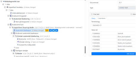
For transparency, some of the issues discussed in the meeting 8th October was:
- How to represent “Sector” of the prostate where the lesions are found?
- There are 24 sectors in Sweden, but not sure they’re internationally recognised. May be not universal.
- Can use Anatomical location archetype, ‘Prostate’ in element Body site name, and use the ‘Specific site’ to carry the sector. Can use SCT codes or other terminologies. Alternatively add the 24 sectors in Sweden in template as a local value set or external terminology, as SCT codes.
- There are several Snomed CT codes regarding regions of the prostate: SNOMED CT - Region of prostate (ihtsdotools.org)
- Or specialise the Anatomical location archetype to something in the line of “Anatomical location – Prostate”? This CLUSTER can be re-used both for Pathological exam and findings, and in treatment (targeted treatment). Further discussion needed.
In which other organs are “Sector” commonly used? These will have similar requirements, and should be handled in the same way.
Example:
In Norway, Snomed CT codes for quadrant for breast are used
- OBS.Imaging exam, element ‘Study date’ says in Descriptipn: “Date/time when the imaging started”.
- How to document “when the image was examined”? Another Event? Or using the Event’s RM element ‘time’?
-
This must be discussed in a broader group to be sure it is consistently used.
-
There is a separate archetype for PI-RADS (soon to be published) that can be used in the anomaly-archetype. .
-
Imaging examination of an anomaly is still under reconstruction.
-
MRI technique details: Suggest to create a separate Cluster with element for ‘magnetic field strength’ as a starting point, instead of using Dose-archetype