Following up on this, we’re currently using the Dosage archetype to record the ordered and administered dosages of such diverse therapeutic procedures as ECT, light therapy, radiation therapy, and hyperbaric oxygen therapy. It’s working well, and I see no reason why the same archetype couldn’t be used to record the dosages from diagnostic imaging procedures ![]()
Hi! I will try to book a meeting to discuss the template. Until then I have yet two other questions:
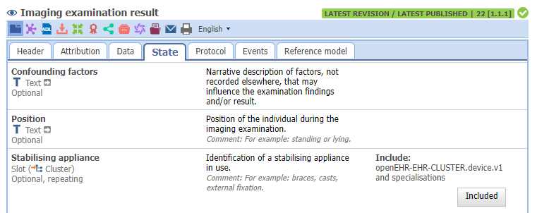
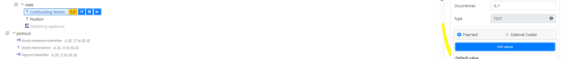
Confounding factors
I would need to capture confounding factors in a structured manner as well as a comment related to each of the confounding factors. I found the Confounding factors attribute in the Imaging examination result-archetype which I guess I could still provide codes for that attribute, but do you have any thoughts on handeling the comment?
Secondary findings
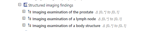
I need to capture findings regarding the prostate (which lots of the ealier discussion relates to), lymph node and bone marrow which I think I handle via multiple structured imaging findings, see below
Above that, I need to capture secondary/incidental findings detected during the MRI study (examination), as in the list below:
- Suspected rectal tumor
- Tumor-suspicious change in the bladder
- Rectal inflammation
- Diverticulosis of sigmoid colon
- Direct/indirect inguinal hernia
- Previous operation with inguinal hernia mesh
- Other
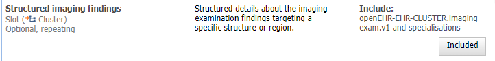
The attribute Structured imaging findings only seems to be suitable for findings targeting specific structures or regions, see below. Any suggestions?
Hi Claudia,
Confounding factors: This element is 0…1, and cannot be repeated. It is a free text element we haven’t thought it would be necessary to document both coded value and free text. We can have a look into that. Meanwhile, do you have any examples of use of multiple codes + descriptions?
Secondary findings: Yes, there will be developed specialisations of the “Imaging examination of a body structure” for specific examinations when there is a concrete use case. The existing ones are only the start. In Norway, we’re also working on prostate cancer, and are happy to collaborate on this.
Incidental findings: We’re still working on how to represent this. There is the “Imaging examination of an anomaly”, which are the one intended for “accidental” findings, but it needs adjustment on the name and in Use section. We’ve just restarted the work on this archetype, and hope to send for another review soon.
@claudiaehr - are the models shared somewhere?
I would like to look into the models (templates, archetypes and terminologies) to compare with our work.
Further down the road I would love to test an import of such data into our EHR system. It would be so great if we could get so far.
Hi, thank you for your replies. I have sent an invite for a meeting next Thuesday and hope that works for you (please let me know if you haven’t gotten an email).
@bna , everything I have been working on regarding openEHR can be found in this branch: regionstockholm/CKM-mirror-via-modellbibliotek at prostate-cancer-radiology (github.com)
@varntzen , the list of confounding factors so far comprises the following (in Swedish and a first translation done by me, has to be validated by a clinician)
- Rörelseartefakter som stör detajlbedömningen (Movement artifacts interfering with detailed assessment)
- Metall/höftledsprotes som stör DWI (Metal/hip prosthesis interfering with DWI)
- Gas i rektum som stör DWI (Gas in rectum interfering with DWI)
- Övrigt (Other)
At a meeting today with among other @heather.leslie , @siljelb, @bna, @varntzen we came to the conclusion that it would be good to have a series of meetings where we model radiology information regarding the prostate together.
If you are interested in joining the work, please let me know in this thread in the near future and I will invite you to the series of meetings.
For transparency, some of the issues discussed in the meeting 8th October was:
- How to represent “Sector” of the prostate where the lesions are found?
- There are 24 sectors in Sweden, but not sure they’re internationally recognised. May be not universal.
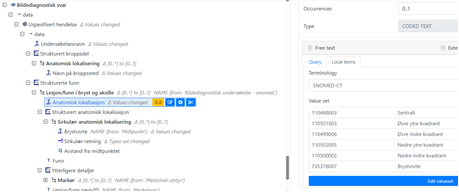
- Can use Anatomical location archetype, ‘Prostate’ in element Body site name, and use the ‘Specific site’ to carry the sector. Can use SCT codes or other terminologies. Alternatively add the 24 sectors in Sweden in template as a local value set or external terminology, as SCT codes.
- There are several Snomed CT codes regarding regions of the prostate: SNOMED CT - Region of prostate (ihtsdotools.org)
- Or specialise the Anatomical location archetype to something in the line of “Anatomical location – Prostate”? This CLUSTER can be re-used both for Pathological exam and findings, and in treatment (targeted treatment). Further discussion needed.
In which other organs are “Sector” commonly used? These will have similar requirements, and should be handled in the same way.
Example:
In Norway, Snomed CT codes for quadrant for breast are used
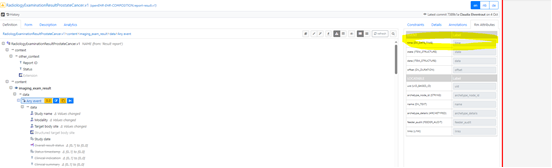
- OBS.Imaging exam, element ‘Study date’ says in Descriptipn: “Date/time when the imaging started”.
- How to document “when the image was examined”? Another Event? Or using the Event’s RM element ‘time’?
-
This must be discussed in a broader group to be sure it is consistently used.
-
There is a separate archetype for PI-RADS (soon to be published) that can be used in the anomaly-archetype. .
-
Imaging examination of an anomaly is still under reconstruction.
-
MRI technique details: Suggest to create a separate Cluster with element for ‘magnetic field strength’ as a starting point, instead of using Dose-archetype